

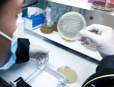

Image analysis can help locate and label candidates that meet a defined rule. The result remains a candidate assessment: appearance alone does not establish species identity or confirm a genetic mutation. A clear record separates what was observed, what the software suggested and what subsequent testing established.
Define the feature you need to observe
Begin with the research question and the reason for comparing colonies. If the task concerns differences in color, size or edge shape, record the comparison group, observation time and classification rule before assigning labels. This makes the selection criteria understandable when someone reviews the images later.
A visible difference between images taken under different conditions may have several explanations. Medium, observation time and imaging conditions belong in the record alongside the feature itself. The American Society for Microbiology's colony morphology protocol describes morphology as a set of observable characteristics and emphasizes recording the growth conditions that influence it.
Use names that describe the observation. “Selected for a different colony appearance” communicates the reason for screening without presenting a genetic conclusion. If a later investigation confirms a biological difference, link that result to the original candidate record rather than replacing the original observation.
Capture images that support comparison
Keep the plate identifier, medium, culture duration, observation time, illumination and imaging settings with each image. Preserve a full-plate view so a close-up can be located again. Check that the relevant area is complete and that reflections, shadows, background color or image compression have not obscured the feature being described.
For size comparisons, record the measurement definition, unit and image calibration. Enlarging a picture on screen does not establish its physical scale. A measurement needs a defensible relationship between image coordinates and the object being measured.
Use a consistent view for comparisons and note when an additional view is needed. An overhead image can show a colony's outline, while a feature involving height requires a view that supports that observation. If the available image does not show a feature reliably, record it as undetermined rather than filling the field from expectation.

Use a repeatable set of observation fields
A structured description makes differences easier to review. The following fields combine visible characteristics with the information needed to find and verify them.
| Observation field | What to record | What can limit interpretation |
|---|---|---|
| Color | Observed color and the label used under the recorded illumination | Light, medium and imaging settings can change its appearance |
| Size | Measurement definition, value, unit and observation time | Scale calibration and growth time must support comparison |
| Shape and margin | Overall outline, visible edge and any uncertainty | Merged growth or an unclear boundary may prevent description |
| Elevation and surface appearance | Features supported by the available view | An overhead photograph may not show height reliably |
| Position | Plate identifier, image reference and target marker | Image labels must remain linked to the physical plate |
| Review status | Software candidate, manually reviewed or unresolved | A candidate label is not biological identification |
ATCC's introduction to microbiology provides descriptions of form, margin, elevation and other colony characteristics. Use a consistent, reviewed vocabulary within a study, and preserve the original observations when terminology is updated.
Review software labels against the original image
Treat automatic labels as a starting point for review. Check whether a candidate includes debris, bubbles, glare or a merged region. A visually convincing marker can still refer to the wrong object, so the original image and the selected region need to remain available together.
Keep unresolved targets in a separate state. When a reviewer changes a label or selection, retain the previous state, the revised state and the reason. Record the classification settings and software version so another reviewer can understand how the candidate list was produced.
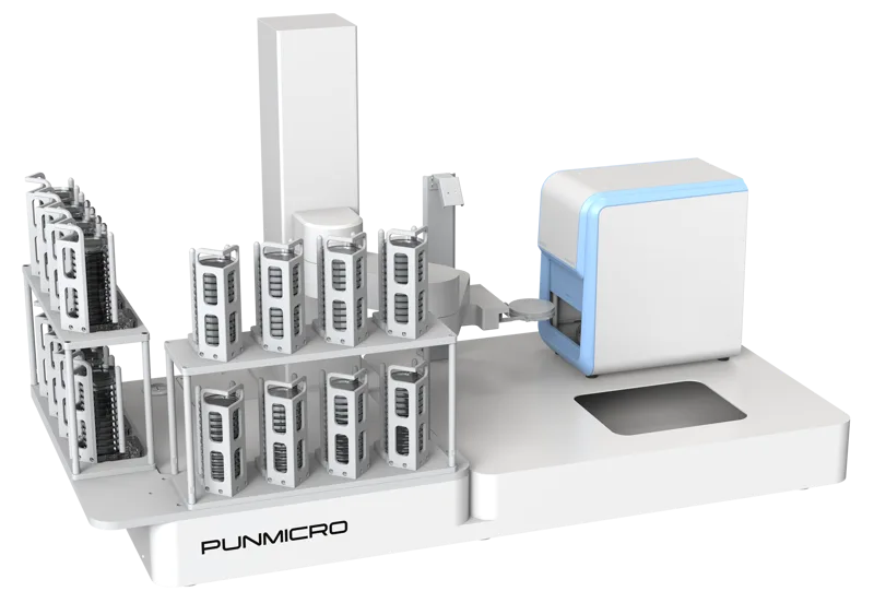
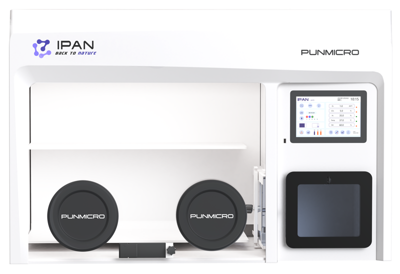
A study's classification rule and an instrument's available functions are separate matters. Features such as circularity, perimeter, texture or automatic category assignment should be checked in the actual software version. When evaluating pun-ICON colony counters, request a demonstration using representative images and the fields you need in the final output.
Keep the link from the image to later work
A candidate list is most useful when every entry points back to a plate, an image and a marked location. Preserve the selection reason and review status as the candidate moves to the next stage. This reduces the chance that an image label and the physical target become disconnected.
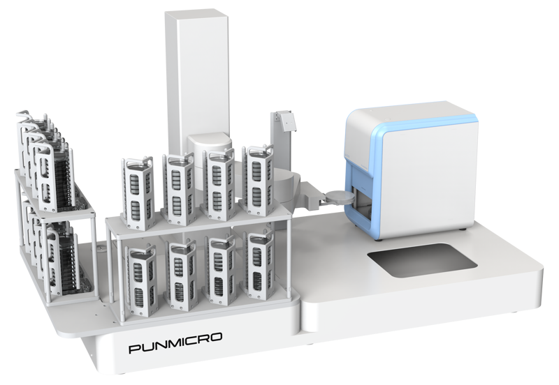
If the next task involves taking material from a colony, review the colony picking workflow and the requirements for a pun-IPICK colony picker. Image counting or classification does not itself establish physical picking capability.
Keep unsuccessful classifications and disagreements as well as selected candidates. These records show where the rules were difficult to apply and help later reviewers judge the scope of the screening decision. For configuration questions, bring the original images, required characteristics and desired output fields to technical support.
Frequently asked questions
Can colony morphology identify a species?
Morphology records visible characteristics and can guide candidate selection. Species identification requires the independent confirmation appropriate to the study; an image label alone is insufficient.
Does enlarging an image make a colony size measurement valid?
No. A size measurement needs a defined unit, measurement method and valid image scale. Screen magnification does not replace calibration.
Can every ICON version calculate the same morphology features?
Check the actual software version and configuration. Request a demonstration and output example for each required feature rather than assuming that a general image-analysis term is available.
References and further reading
- ASM Colony Morphology Protocol · Read the official protocol; supports the visible-characteristic framework and the need to record growth conditions. No culture procedure reproduced.
- ATCC Introduction to Microbiology · Read official search-return content describing shape, margin, elevation, size and optical appearance; terminology only.