

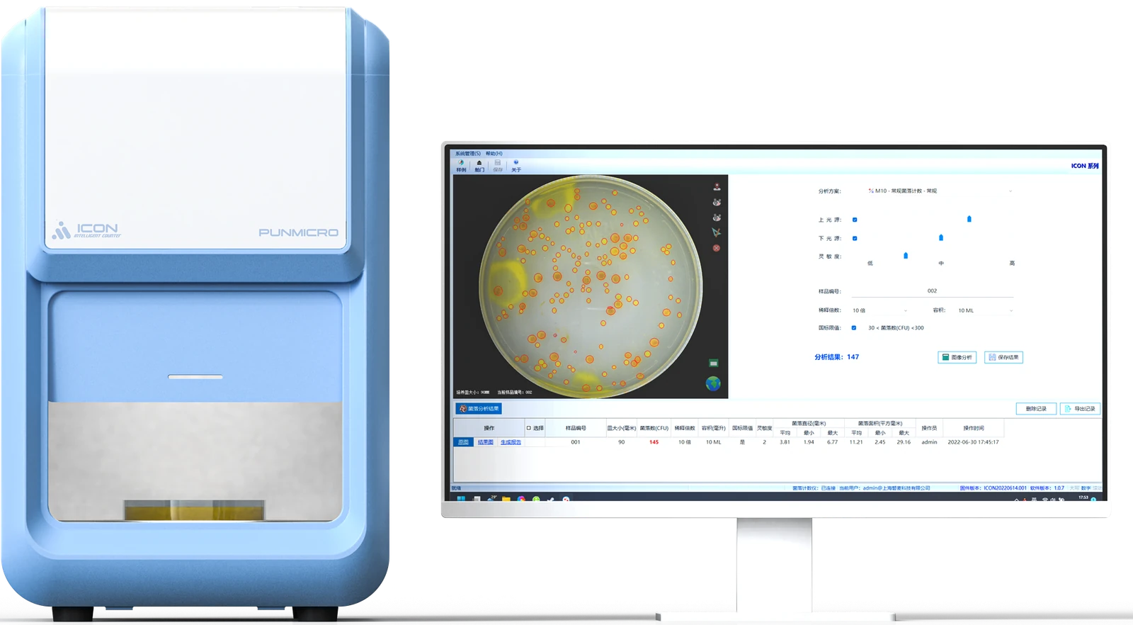
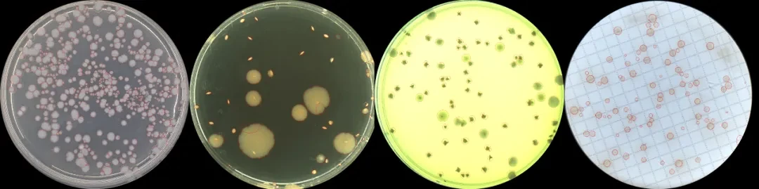

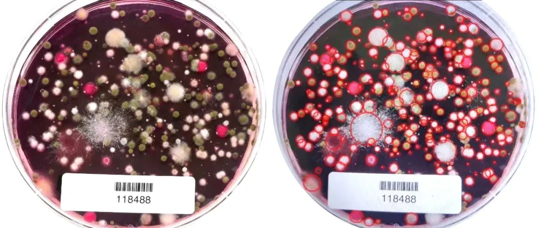



A useful counting record connects the plate's identity to its original image, the reviewed markers and the reported result. This framework keeps that connection intact without applying one counting range or image setting to every test.
Confirm the plate's identity and condition
Before reading, check the sample identifier, dilution level, inoculation method, culture record and test being performed. Similar-looking plates may belong to different dilutions or parallel preparations of the same sample. Resolve identity questions before a count is attached to a result.
Inspect the medium and plate for damage, contamination, condensation, foreign material and extensive merged growth. The acceptable counting range and treatment of abnormal plates come from the applicable method. A fixed range should not be copied across unrelated tests.
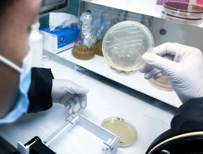
Position the plate securely with the intended area fully in view. Decisions about removing a lid, managing condensation or acquiring another image should follow the instrument instructions and laboratory procedure. A clearer photograph is not a sufficient reason to change the sample's condition outside that procedure.
Inspect the image before accepting its count
Check the center and edge for focus, uneven lighting, reflection, shadow, obstruction and color changes. Small colonies require discernible information in the original image; enlarging a display cannot supply detail that was never captured.
If the target and background cannot be separated reliably, review the plate condition and imaging setup before continuing. The question is whether the image supports a defensible reading, not whether the software can produce a number.
Image analysis may involve background processing, target segmentation, feature recognition and counting. Implementation differs between systems. A general explanation of thresholding, watershed segmentation or morphological processing should not be treated as a description of the internal algorithm in a particular ICON version.

Compare detection markers with visible targets
Read the marked image against the original. Check both detected targets and areas where a target may have been missed.
| Image feature | Review question | Record to keep |
|---|---|---|
| Bubbles, particles or scratches | Has a non-colony object been counted? | Location, reason for the decision and any correction |
| Small or low-contrast colonies | Has a visible target been omitted? | Reviewed image detail and the basis for including it |
| Touching or merged growth | Has the image been split too far, or have distinguishable targets been combined? | Method-based reading decision and its limitations |
| Plate edge | Has reflection created a marker, or has part of the counting area been cropped? | Actual region counted and reasons for exclusions |
| Colonies of different colors | Are these objects within the test's target definition? | Classification rule and confirmation status |
When a marker is added or removed, retain the result before and after the change and the reason for it. For repeat readings, use the agreed method and settings. Record any setting change rather than repeatedly adjusting the image until the total approaches an expected value.
An unresolved target can remain unresolved in the record. A precise-looking integer does not make a contaminated, extensively spreading or otherwise uninterpretable plate acceptable.
Separate the plate count from the reported concentration
The number of colonies on one plate and the concentration reported for the original sample are different fields. Concentration calculations also require the dilution relationship, inoculated volume and, where relevant, the original sample's mass or volume basis.
The method determines how results from multiple dilution levels are combined and how abnormal plates are handled. See the CFU calculation guide for the distinction between the count, calculation inputs and reporting unit. Keep the full calculation linked to the plate record.
| Record group | Information to retain |
|---|---|
| Identity | Sample ID, plate ID, test and method version |
| Image evidence | Original image, marked image and counting region |
| Reading | Initial automatic count, reviewed count and changes with reasons |
| Calculation inputs | Dilution, inoculated volume and applicable sample basis |
| Result status | Reported unit, method-based interpretation and unresolved limitations |
For a plate affected by contamination, excessive spreading, indistinguishable targets or a result outside the method's scope, preserve the abnormal status and its disposition. Do not replace the qualification with a normal-looking count.
Check repeatability without confusing sources of variation
Repeated analysis of the same plate or an approved reference image can help examine consistency under a predefined plan. Independent review by another reader may also be appropriate. Record the image, settings and reading rules so that a difference can be traced.
Keep three comparisons separate: repeated instrument readings of the same material, different readers' decisions, and results from separate parallel plates. Parallel plates can also reflect biological and preparation differences. Their variation cannot all be assigned to the counter.
When results differ, inspect imaging, sample condition, settings and interpretation rules, then record the decision and its basis. For a structured equipment comparison, see manual versus automated colony counting.
Frequently asked questions
Can the same counting range be used for every method?
No. Use the range and abnormal-plate rules specified for the test. The counter assists the reading; it does not establish the method's acceptance conditions.
Should an unclear target always be split into separate colonies?
No. Preserve the image and apply the relevant reading rule. Where boundaries cannot be interpreted reliably, retain the limitation or abnormal status rather than forcing a count.
What should be saved after a manual correction?
Keep the original image, marked result, count before and after the change, reason for the correction and its link to the sample and plate. The final report should remain traceable to that record.
For equipment selection, visit pun-ICON automatic colony counters. Model, maintenance and software-version questions can be directed to technical support.