



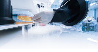
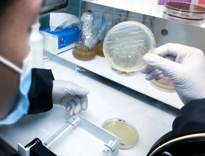

The same image workflow may be relevant to several kinds of plates, but their samples and reporting requirements remain distinct. Establish what was collected and what the test is intended to measure before deciding which objects to count.
Distinguish the sample from the counting task
Drinking water, surface water, wastewater, air and surfaces do not share one sampling method or reporting limit. Water testing may involve membrane filtration or plate culture, while air and surface samples have their own collection approaches.
Keep those differences visible in the records. A result derived from one sample basis should not be presented with the concentration unit or interpretation used for another task.
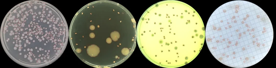
For a membrane-filter plate, assess the membrane color, grid, background particles and target appearance after culture. Contact plates and other dishes also need a check of their dimensions, surface structure and edges.
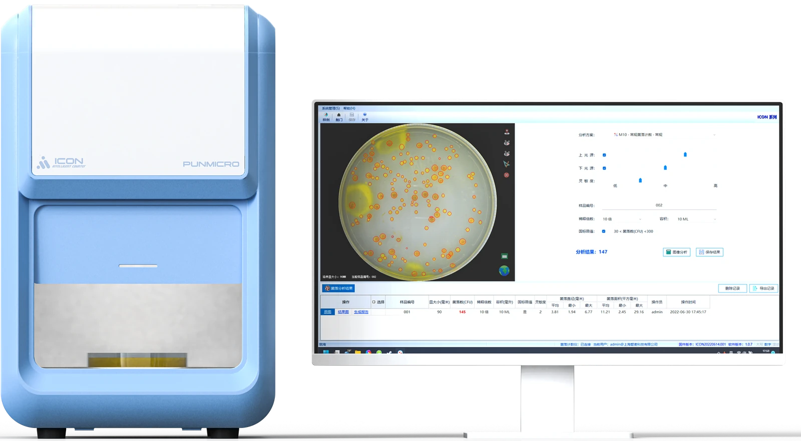
When evaluating ICON, confirm membrane or contact-plate suitability using the actual sample, consumable and software configuration. A general reference to a plate category does not establish that every format or method has been evaluated.
Identify the interference before correcting a marker
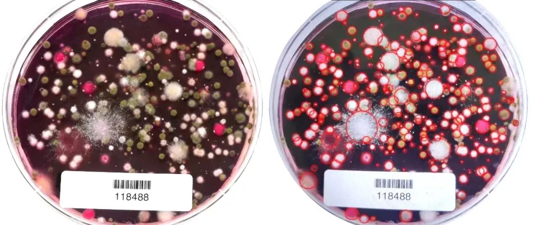
Environmental samples can contain objects and backgrounds that complicate image interpretation. Compare the original image with the markers and record the reason for a change.
| Image problem | Possible counting effect | Review focus |
|---|---|---|
| Particles or deposits from a water sample | A non-colony object may enter the count | Appearance, location and relationship to the sample background |
| Very small or faint colonies | A visible target may be missed | Original image detail and contrast |
| Grids, scratches or reflections | False targets or obscured boundaries | Correspondence between markers and actual outlines |
| Spreading or extensively merged growth | Separate targets may not be distinguishable | Treatment of abnormal plates under the method |
| Several colors or forms | The total and the intended target category may be confused | The target definition for this test |
An image algorithm cannot establish a biological boundary that is no longer discernible in the image. Where growth is extensively merged or the plate cannot be read reliably, use the applicable method's handling rules and retain the limitation. Do not force the image into an arbitrary number of separate objects.
A useful correction record states what was visible, where the marker differed and why the reading changed. It also preserves targets that remain uncertain. This makes the review understandable when someone returns to the record later.

Preserve the link from sampling to the reported count
For each plate, retain the sampling location, sample type, plate ID, test, dilution relationship, inoculated or filtered amount and culture information. After analysis, save the original image, marked image, initial automatic value, reviewed value and abnormal-result explanation.
The reported concentration requires the inputs and units appropriate to the method. Keep the plate reading and any subsequent calculation distinguishable so that the result can be traced back without guessing which volume or dilution was used.
| Record layer | Information to connect |
|---|---|
| Sampling | Location, sample type and sample identifier |
| Preparation and culture | Dilution, inoculated or filtered amount, method and culture record |
| Plate reading | Plate ID, original image, counting region and marked image |
| Review | Initial value, changes, reviewed value and unresolved observations |
| Report | Calculation inputs, required unit and the method-based interpretation |
For batch work, distinguish dilutions and parallel samples from the same location. Their names may look similar while referring to different plates and preparation records.
Before comparing parallel results, check sampling, preparation and culture conditions. Differences between separate plates cannot all be attributed to the counter. Keep the evidence for those stages alongside the image review.
Treat color and fluorescence as observations
Some environmental investigations include observations of colony color or fluorescence, including images of colonies suspected to be Legionella. Those observations need the confirmation required by the applicable method. Color or fluorescence alone does not establish species identity.
If a proposed counting configuration includes ultraviolet or fluorescence-related imaging, confirm its documented wavelength, illumination conditions, compatible models and any implications for subsequent sample use. The presence of a fluorescent image does not demonstrate that a particular instrument supports the assay or preserves every sample property.
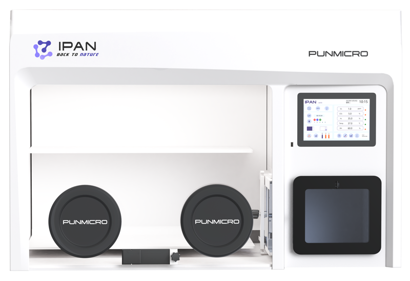
Keep imaging, cultivation and environmental-control equipment separate in the specification. ICON's image-counting role does not establish gas-control capabilities, biological containment or unconditional protection of sample viability. These questions need their own equipment and method evidence.
This page addresses image interpretation and record relationships; organism-specific test conditions remain with the relevant method and responsible laboratory personnel.
Evaluate the workflow with routine and difficult samples
Use a plate set that includes ordinary samples and the high-interference backgrounds encountered in practice. Check target detection, manual changes and saved reports together.
For each sample group, record which types could be handled, how much review was needed and which plates could not be resolved. Retain the reason for an exclusion or limitation instead of removing the example from the assessment.
Confirm the actual consumable, counting region and software configuration used. This turns a general compatibility discussion into an evaluation of the plates your laboratory needs to read. The pun-ICON product guide covers these configuration questions.
For the common image-review sequence, see how to count colonies on agar plates. Maintenance, lighting and image-quality concerns can be referred to technical support with the relevant model and image records.
Frequently asked questions
Can one reporting unit be used for every environmental sample?
No. The sample basis, collection approach and test method determine the required inputs and units. Water, air and surface tasks should retain their separate context.
Does a fluorescent colony image identify the organism?
No. Color or fluorescence provides an observation. Identity requires the confirmation specified by the applicable method.
Can software resolve every area of merged growth?
No. Where boundaries cannot be read reliably, preserve the limitation and follow the method's abnormal-plate rules. A numerical output does not remove that uncertainty.